Explore
 Validate
 Learn
LearnNBP2-34638-0.1 mg
antibody from Novus Biologicals
Targeting: PMEL
D12S53E, gp100, Pmel17, SI, SIL, SILV
 Western blot
 Immunohistochemistry
Antibody data
- Antibody Data
- Antigen structure
- References [0]
- Comments [0]
- Validations
- Immunohistochemistry [3]
- Flow cytometry [2]
Submit
Validation data
Reference
Comment
Report error
- Product number
- NBP2-34638-0.1 mg - Provider product page

- Provider
- Novus Biologicals
- Product name
- Mouse Monoclonal PMEL17/SILV Antibody
- Antibody type
- Monoclonal
- Description
- Protein A or G purified. By immunohistochemistry, it specifically recognizes a protein in melanocytes and melanomas. This MAb reacts with junctional and blue nevus cells and variably with fetal and neonatal melanocytes. Intradermal nevi, normal adult melanocytes, and non-melanocytic cells are negative. It does not stain tumor cells of epithelial, lymphoid, glial, or mesenchymal origin. Metastatic amelanotic melanoma can often be confused with a variety of poorly differentiated carcinomas, large cell lymphomas, and sarcomas using H & E stains alone. It is also difficult to differentiate melanoma from spindle cell carcinomas and various types of mesenchymal neoplasms. This MAb stains fetal and neonatal melanocytes, junctional and blue nevus cells, and malignant melanoma. This MAb also stains Angiomyolipoma (PEComa).
- Reactivity
- Human, Rat, Canine
- Host
- Mouse
- Isotype
- IgG
- Vial size
- 0.1 mg
- Concentration
- 1.0 mg/ml
- Storage
- Store at 4C short term. Aliquot and store at -20C long term. Avoid freeze-thaw cycles.
No comments: Submit comment
Supportive validation
- Submitted by
- Novus Biologicals (provider)
- Main image
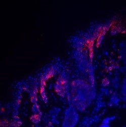
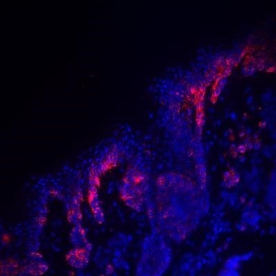
- Experimental details
- Immunohistochemistry: PMEL17/SILV Antibody (HMB45) - Azide and BSA Free [NBP2-34638] - PMEL17 (red) was detected in human skin (melanoma) using PMEL17-PE antibody (1:200) in PBS for 1 hour. Nuclei were stained with DAPI (blue). Image from a verified customer review. Image using the PE format of this antibody.
- Submitted by
- Novus Biologicals (provider)
- Main image
- Experimental details
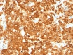
- Immunohistochemistry-Paraffin: PMEL17/SILV Antibody (HMB45) - Azide and BSA Free [NBP2-34638] - Formalin-fixed, paraffin-embedded human testis stained with gp100/Melanosome Monoclonal Antibody (HMB45).

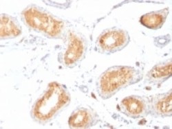
- Submitted by
- Novus Biologicals (provider)
- Main image
- Experimental details
- Immunohistochemistry-Paraffin: PMEL17/SILV Antibody (HMB45) - Azide and BSA Free [NBP2-34638] - Formalin-fixed, paraffin-embedded human melanoma stained with gp100/Melanosome Monoclonal Antibody (HMB45).

Supportive validation
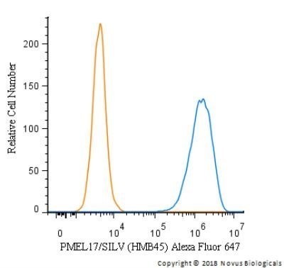
- Submitted by
- Novus Biologicals (provider)
- Main image
- Experimental details
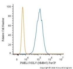
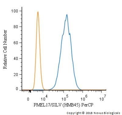
- Flow (Intracellular): PMEL17/SILV Antibody (HMB45) - Azide and BSA Free [NBP2-34638] - An intracellular stain was performed on SK-MEL-28 cells with PMEL17/SILV Antibody (HMB45) NBP2-34638PCP (blue) and a matched isotype control (orange). Cells were fixed with 4% PFA and then permeabilized with 0.1% saponin. Cells were incubated in an antibody dilution of 5 ug/mL for 30 minutes at room temperature. Both antibodies were conjugated to PerCP. Image using the Azide and BSA Free form of this antibody.
- Submitted by
- Novus Biologicals (provider)
- Main image
- Experimental details
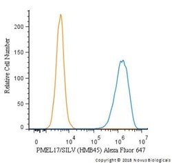
- Flow (Intracellular): PMEL17/SILV Antibody (HMB45) - Azide and BSA Free [NBP2-34638] - An intracellular stain was performed on SK-MEL-28 cells with PMEL17/SILV Antibody [HMB45] NBP2-34638B (blue) and a matched isotype control (orange). Both antibodies were conjugated to Biotin. Cells were fixed with 4% PFA and then permeabilized with 0.1% saponin. Cells were incubated in an antibody dilution of 2.5 ug/mL for 30 minutes at room temperature, followed by Streptavidin - R-Phycoerythrin Protein (2012-1000, Novus Biologicals). Image using the Biotin format of this antibody.